Showing posts with label teaching tips. Show all posts

Wednesday, March 18, 2020

COVID-19 Scramble: Adapting to Remote Learning, Suddenly

Well, friends, I'll bet most of us didn't see this pandemic coming. Not to the extent of impact it's had on the teaching and learning of anatomy and physiology. But it's here and we're going to roll with the punches and deliver a positive and productive learning experience for our students, right?

To help us all get ourselves organized and on the right track as we adapt to suddenly having to move our face-to-face course homes to a remote venue out there in the vast expanse of space, I've assembled a few "get started" resources to help you adapt.


Quickly Moving to Remote Delivery—The Musical

This is an "emergency" bonus episode of my podcast (The A&P Professor). The brief audio presentation presents nineteen tips on how to get started. And there are three A&P songs from my friend and fellow A&P teacher, Greg Crowther. For a sing-along! We could all use a light-hearted sing-along about sodium ions right now, am I right?

Go to theAPprofessor.org/64b for an audio player plus a lot of links and other resources. Or subscribe in your favorite podcast player and look for Episode 64b. If you subscribe, you can access the links and images in the shows notes of each episode right there in your podcast player. And you'll also automatically get more episodes with tips on how to cope with the current situation—and so much more

There is also an earlier episode called Mid-Winter Winterizing of Our Courses meant to help prepare us before this all flooded in upon us. Many of these prep tips are still useful, even as the metaphorical flood waters continue to rise all around us. Go to theAPprofessor.org/63b

TEACH Instructor Resources for Anatomy & Physiology

This little gem has been there all along! Really. In your Evolve (Instructor) Resources for The Human Body in Health & Disease, 7th Edition  TEACH has all kinds of tips and strategies that can be used to generate ideas for learning activities that can be adapted for remote teaching.

Included in TEACH are:
  • Lesson Plans
  • Student Handouts
  • PowerPoint Slides
  • Pretest Questions and Pretest Answers


If you don't have an Evolve account, then just go to evolve.elsevier.com and click on Sign In in the upper right, then click on Create Account near the bottom of the form that pops up. Make sure you apply for a faculty account. It will take a day or so to verify your faculty status.


In the search box on the home page, or when you click Catalog at the top, type in Evolve Resources for The Human Body in Health & Disease, 7th Edition, then request access. The TEACH resources will be listed in the Instructor Resources tab.


But wait! There's more...

Besides the TEACH resources and Teaching Tips, there are all kinds of other resources in your  Evolve (Instructor) Resources for The Human Body in Health & Disease, 7th Edition:

  • Audience Response Questions—meant for "clicker" systems in a classroom, they can be easily adapted for online presentations to spark student thinking during an online "lecture" or demonstration.
  • Image Collection—contains (labeled and unlabeled) jpeg and PowerPoint versions of each image from the textbook.
  • Test Bank—can be a great resource for quickly constructing online quizzes and reviews. Consider using them for Testing-as-Teaching, a type of retrieval practice mentioned later in this post.

The A&P Professor

I already mentioned The A&P Professor podcast, but there is lot more there for you than those "emergency" bonus episodes. It's worth exploring the whole list for practical tips and advice as you move to remote teaching.

The great thing about podcast episodes is that you can listen to them while you are wiping down the surfaces in your home, making your family's meals, and rearranging your stacks of toilet paper.

Here are a few selected topics to start with:


Besides the podcast, The A&P Professor website includes other resources, such as online seminars for teaching anatomy and physiology. Here are a couple that may be helpful as this time:

Free eText

VitalSource and Elsevier have partnered to provide eText access to students. To assist students at disrupted semester-calendar schools who are losing access to course materials due to COVID-19 campus closures, VitalSource has been joined by Elsevier to offer free access to etexts to students whose classes have moved online from March 16 through May 25, 2020. Students will be able to access the expansive catalog of eTexts from participating publishers through the VitalSource Bookshelf app effective immediately.


Besides this being useful to students who are "stuck without" their textbooks, it can offer additional opportunities to connect with students in a remote environment. 

For example, something that I do in an online course I teach is let my students "subscribe" to the highlights and notes in my own copy of the eText version of the textbook. 

I can mark areas of particular importance, add commentary on what they should be looking at in a section or illustration, and clarify concepts that commonly challenge student learning. This could be particularly useful in "holding the hand" of a confused and dazed student who is trying to adapt to a new learning environment.

Coloring Book

I've been seeing a lot of advice to folks in general, telling them to sit around as a family and color in their coloring books. That makes a lot of sense in terms of diffusing stress and connecting in a positive way with those with whom we are house-bound. 

Why not suggest to students to do that, but use Mosby's Anatomy & Physiology Coloring Book as one of their coloring books? 

In a post from my blog The A&P Student, I recommend coloring as a way to study anatomy and physiology in a new and fun way—that also calms the nerves. So their nerves will become calm as they study their nerves! Check out Coloring Books Are Powerful Study Tools (And They Help Manage Stress).


Okay, whew! That's enough for now, eh? Don't hesitate to reach out if I can be of help to you. And don't forget your Elsevier Education Solutions Consultant!

Thursday, February 28, 2019

Celebrating Women's History Month in The Human Body in Health & Disease

March is Women's History Month and as you consider ways to celebrate the role of women in the human sciences, why not start in your textbook?

The Human Body in Health & Disease has several descriptions of the contributions of women who have made important strides in understanding human structure and function.

Elaborating on these stories yourself  is one way to celebrate Women's History Month. Another is to assign students to find more information about one or more of these women. Perhaps they could present this information to their class in the form of a blog or wiki entry, a poster or handout, or other creative media.

For example, the Growth & Development chapter includes a mention of Rita Levi-Montalcini's contributions to understanding the development of the nervous system.

In the Reproduction chapter, we recognize the role of Virginia Johnson in early attempts to understand human sexuality.

In the Chemistry chapter, we highlight the role of Rosalind Franklin in understanding the structure of DNA.

Why not start a conversation in your course today about the role of women in understanding the  the human body in health and disease?

Thursday, February 7, 2019

African-American History Month in The Human Body in Health & Disease

If you are thinking about ways to celebrate the roles of African-Americans in scientific discoveries related to human biology as part of African-American History Month (also known as Black History Month), then you can start in your textbook.

The Human Body in Health & Disease has several built-in resources to jump-start a conversation.

For example, the Blood chapter includes a boxed sidebar that highlights the contributions of Charles Richard Drew to hematology—particularly in the establishment of blood banks.

In the Nutrition & Metabolism chapter, there's another boxed sidebar that mentions the role of George Washington Carver in the rise of food science. 

Just these two snippets can be a conversation starter in your course. Consider asking your students to contribute more—perhaps as a project or other assignment.

You might also want to check out Black History Month: Celebrating Blacks in Science, Promoting Diversity in STEM to stimulate some ideas for your course.

Wednesday, May 17, 2017

Help Students Master the Language of Science & Medicine

You can't really learn and experience what's going on in your world until you can use the language. In a beginning  course, most students don't come in as native speakers of scientific and medical terminology.

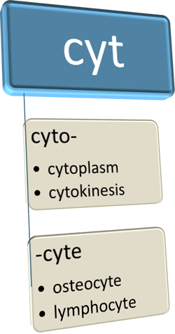
Even if they know what roots, suffixes, and prefixes are, most of them don't know the literal meanings of "meta," "juxta," "reno," or "cyto." If they pick up these word parts as they learn, however, they can quickly get comfortable with the language of science and medicine—and really ramp up their learning of core concepts.

In The Human Body in Health & Disease, we have woven language learning into every chapter so that students can more efficiently master the language of science and medicine.

Nearly every anatomy and physiology and patho teacher I know mentions word parts frequently in class discussions—especially when introducing the more convoluted terms that represent important concepts.

Besides breaking down impossibly long words into easily consumed, bite-sized pieces, it's also a stealthy strategy to "sneak in" some language learning. Without having to "put it on the test" we can introduce word parts— and how they are used to construct phrase-like terms—in a way that allows natural language learning. Students often don't even realize that our repeated mentioning of the meaning of common word parts is adding to their mental lexicon. Soon they start recognizing these word parts on their own.

When students know what common word parts mean, they start using them as mnemonic tools (memory aids). They find themselves using the word parts as clues to remember the actual working definition of the term—and the essence of the concept that the term represents.

In The Human Body in Health & Disease, we support this widely used classroom approach in several ways. A central strategy is our inclusion of word parts in the chapter word lists.

Previewing unfamiliar words before reading a chapter helps to get those words into the reader's mental lexicon more quickly—and how that, in turn, improves reading comprehension. Because we also include word parts in the chapter word lists, scanning these lists naturally builds competence in scientific and clinical language.

A widespread and effective strategy in teaching reading skills—from elementary school to advanced courses in college reading efficiency—is often called word study. In a nutshell, word-study instruction encourages readers to strengthen their recognition of word patterns as first step, rather than simply memorizing new words as they encounter them. This is based on the fact that we read words and phrases as a whole. It's one of many brain-based strategies of learning that translate current concepts of neuroscience into practical strategies.

Incorporating word parts in the chapter word list, where they can be scanned before reading the chapter, offers the opportunity for readers of The Human Body in Health & Disease to make the word study method part of their overall reading strategy. By building pattern-recognition skills, readers can better get new terms into their mental lexicon and thus be able to read them without stumbling and thus learn concepts more efficiently.

But wait! There's more! In The Human Body in Health & Diseasewe provide even more built-in tools to help students gain skill in using their new language. In an upcoming post, I'll point these out.


Article content adapted from Anatomy & Physiology

Wednesday, May 3, 2017

Help Students Integrate Concepts with CONNECT IT!

New to The Human Body in Health & Disease is a supplemental feature called Connect It!. First introduced in our larger textbook Anatomy & Physiology as A&P Connect, it was a logical evolution of sidebars and boxed essays as we progress through a new age of digital learning tools.

We've always heard from students how much they like boxed essays in the print version of our textbooks. The more our users explored, the more interested in—and enthused about—anatomy and physiology they become.

Putting additional topics of interest on a digital platform seemed to be a great way to better satisfy the natural curiosity of students—and let students get a deeper understanding of concepts by exploring clinical applications and current research trends.

What we soon found out was that branching out into brief sidebars that reside on the digital Evolve platform, we had the freedom to add diagrams, anatomical art, medical images, and micrographs that we simply didn't have room to add to the textbook proper.  Because most concepts of human structure, function, and pathophysiology, benefit by such visual representations, students have welcomed this evolution of the traditional sidebar.

Because Connect It! articles are not located in a particular spot in a print book, they can be easily referenced in any part of the book.  We leveraged this strategy to help student better integrate the concepts they are learning.

For example, the new Connect It! article on the human microbiome not only explains an emerging central concept of human biology—it also allows us to apply the concept to almost every system of the body. The article itself integrates many organ systems in its approach, so when it is reviewed in different chapters of the book, new and deeper understanding of how the functions of the body are integrated emerges.

Repeated mention of an integrating topic also reinforces the understanding of the connectedness of human structure and function. Such integration also helps deepen the understanding of general principles of pathophysiology by applying them to several different disorders.

Another example is our Connect It! article Protective Strategies of the Respiratory Tract, which helps integrate concepts across discussions of respiratory structure and function, as well as immunity. Likewise, the article Medical Imaging of the Body links concepts that appear in several different chapters.

The fact that the Connect It! articles reside outside the textbook proper also gives the instructor some flexibility. For example, you might pick a few of the topics for special emphasis in your course and provide one or other of the Connect It! articles as a lecture/discussion handout—perhaps as a resource when solving a case study or as part of a set of class notes/study guide. Simply copy and paste from the article to your course materials! Another option is to mention an article by name in your syllabus or course outline.

You may also consider using some of the diagrams or other images from the Connect It!  in your teaching slides or other teaching materials.

I'm very excited about this intentional use of our Connect It! articles in helping students integrate concepts of structure, function, and pathophysiology to achieve a deeper understanding of human biology for the health professions. And professors will appreciate the role of this new feature in elevating student enthusiasm and motivation!


Thursday, September 29, 2016

Here's the Scoop!

This blog is a way for me to communicate with users and potential adopters of my anatomy and physiology textbook, The Human Body in Health & Disease.

In coming months, look for brief articles that explain the learning theory behind our textbook's features and design. I'll alert you to changes to each new edition—and the rationale behind them. There will be behind-the-scenes information about how the book is created. I'll describe best practices in using the textbook in your course, with your students.

To start the ball rolling, I'll tell you about that crazy image on the cover of the new 7th edition due out in February 2017.

It's a scanning electron micrograph (SEM) of methicillin-resistant Staphylococcus aureus—often called MRSA (MER-sah). The image has false color added, rendering the MRSA organisms as a golden yellow.  The large, reddish, blob-like structure is the remains of a dead human neutrophil.

The image was selected for several reasons. We wanted a dramatic but "real" photo as the main image.  We looked for something from the human body, but also were interested in reflecting the "disease" and related clinical aspects of the textbook's content.  MRSA is an important pathogenic bacterium in human medicine, and this micrograph shows the damage to human cells that it can wreak in the human body.

An interesting classroom or online discussion exercise to ask students what it looks like to them. When the answer is discovered, ask students what they already know about bacterial infections—and MRSA in particular.  This may serve as part of your first-day class activities to generate interest and interaction among a new group of learners!

Photo: NIAID